

 8 hours ago
5
PROTECT YOUR DNA WITH QUANTUM TECHNOLOGY
Orgo-Life the new way to the future Advertising by AdpathwayA retrospective neuroimaging investigation has demonstrated that spontaneous intracranial hypotension induces quantifiable, region-specific mechanical strain across the human brain parenchyma, identifying pronounced tissue deformation within fronto-limbic and temporomesial networks that persists independently of gross volumetric shifts. Analyzing ninety-six patients who underwent surgical occlusion of spinal cerebrospinal fluid leaks alongside sixty-one healthy control subjects, the research team employed artificial intelligence-assisted diffeomorphic registration of volumetric magnetic resonance imaging scans to establish voxel-wise displacement fields across one hundred fifty-five segmented brain regions. The resulting biomechanical models revealed that while fluid-filled ventricles and venous sinuses exhibited substantial perioperative volumetric expansion and contraction, brain parenchyma within the temporal poles, basal forebrain, hippocampus, and orbitofrontal cortex underwent marked distortional shearing. These quantitative findings demonstrate that downward brain displacement in spinal cerebrospinal fluid leaks exerts localized mechanical stress upon neural networks governing cognitive and behavioral regulation.
Spontaneous intracranial hypotension represents an underdiagnosed neurological condition precipitated by structural cerebrospinal fluid leaks along the spinal column. Although the hallmark clinical symptom remains debilitating orthostatic headache, prolonged cerebrospinal fluid loss can generate an expansive spectrum of neurological disturbances, including auditory tinnitus, diplopia, executive dysfunction, and marked cognitive alterations. In advanced presentations, patients may manifest profound neuropsychiatric symptoms and behavioral disturbances that closely mimic the behavioral variant of frontotemporal dementia, a clinical convergence that has given rise to the diagnostic concepts of brain sagging dementia and spinal dementia. Historically, understanding these complex neuropsychiatric presentations has been hindered by a lack of objective tools capable of quantifying physical stress within deep cerebral structures. While prompt clinical recovery following surgical closure of spinal defects has long pointed toward reversible functional disruption rather than permanent tissue loss, the underlying mechanical pathways linking fluid depletion to cognitive impairment have remained uncharacterized.
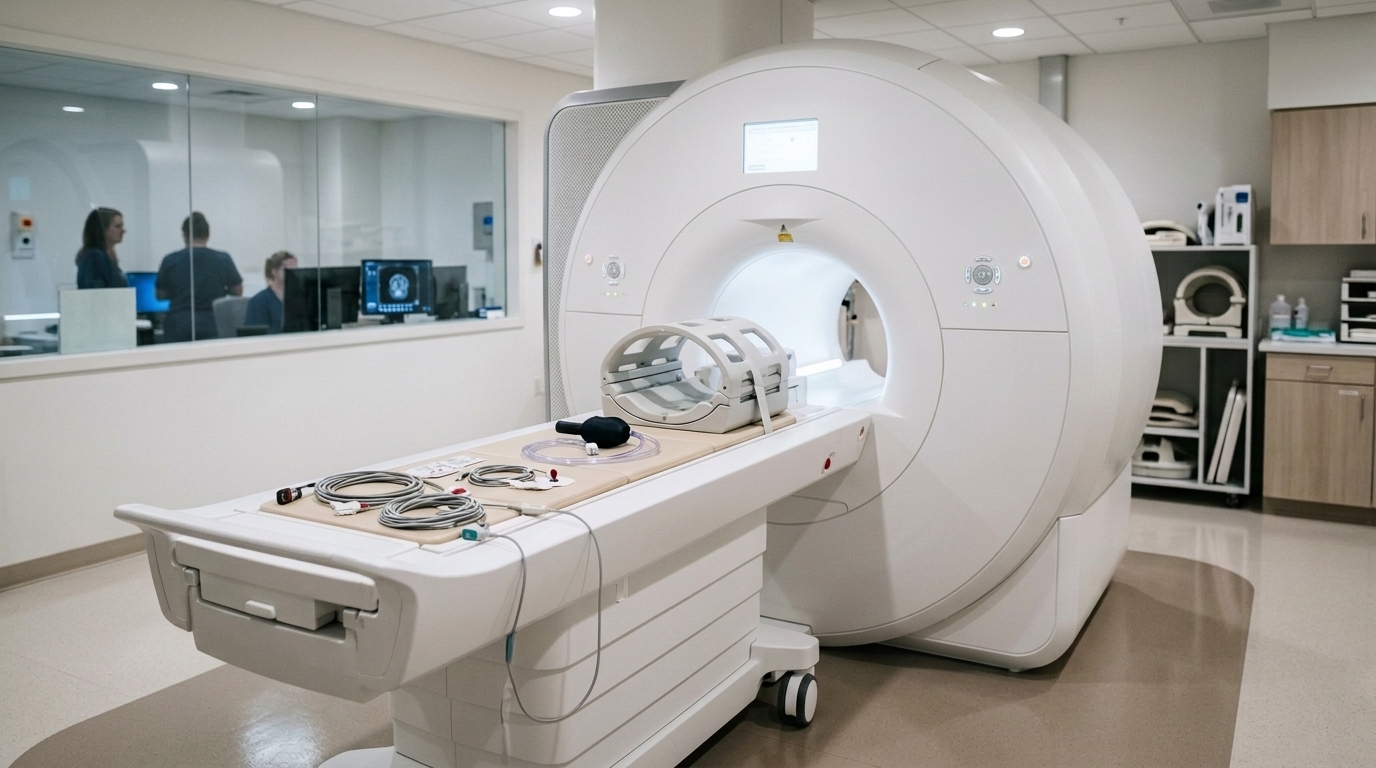
Diagnostic evaluation and therapeutic follow-up in spontaneous intracranial hypotension rely predominantly on structural magnetic resonance imaging to detect characteristic qualitative signs collectively known as brain sagging. Established clinical scoring systems, such as the score introduced by Dobrocky and colleagues, systematically evaluate morphological hallmarks including the effacement of basal cerebrospinal fluid cisterns, venous engorgement, diffuse pachymeningeal enhancement, and subdural fluid collections. Nevertheless, these conventional radiological assessments remain fundamentally descriptive and interpret brain sagging as a uniform downward displacement of intracranial structures within the rigid cranial vault. Such assessments cannot separate uniform physical displacement from localized parenchymal deformation, nor can they quantify mechanical stresses experienced by specific anatomical structures. In biological tissues, downward displacement induces both local volume changes and distortional shearing forces that vary according to anatomical geometry. Characterizing these mechanical forces requires automated, reproducible computational methods capable of measuring voxel-wise deformation fields across interconnected cerebral networks.
To quantitatively investigate parenchymal deformation, investigators conducted a single-center retrospective study approved by the institutional ethics committee under reference identifiers 22-1512-S1-retro and 24-1296-S1-retro, adhering strictly to good clinical practice standards. The patient cohort comprised individuals evaluated at a tertiary referral center who satisfied the Third Edition of the International Classification of Headache Disorders criteria for spontaneous intracranial hypotension and underwent surgical repair of spinal cerebrospinal fluid leaks between April 2018 and December 2024. Eligible participants possessed isotropic contrast-enhanced T1-weighted magnetic resonance images with a minimum spatial resolution of one millimeter acquired within twelve months before surgery and between one and twelve months postoperatively. The final analysis included ninety-six patients with a mean age of 43.4 years, of whom 62.5 percent were female. Median intervals spanned six days from preoperative imaging to surgery, ninety-eight days from surgery to postoperative imaging, and one hundred twelve days between baseline and follow-up scans.
To establish an empirical baseline for registration variability and physiological scanner noise, the researchers utilized longitudinal imaging data from the Beijing Normal University 2 dataset, curated by the Consortium for Reliability and Reproducibility. This control cohort comprised sixty-one healthy adults aged between nineteen and twenty-three years who underwent two unenhanced T1-weighted magnetic resonance examinations separated by 103 to 189 days. Patients presenting with subdural hygromas, chronic hematomas, or significant metallic susceptibility artifacts were systematically excluded from the surgical cohort to prevent massive fluid shifts from distorting subtle parenchymal strain calculations. The authors noted that the control group, being younger and scanned on different systems without gadolinium contrast, provided a conservative standard of comparison, as technical differences between scanners typically elevate apparent background deformation. Despite these scanner differences, applying the computational workflow to healthy controls yielded near-zero baseline deformation, confirming that patient distortions reflected true pathological processes.
The image processing pipeline converted raw DICOM data into standardized NIfTI formats before executing automated anatomical segmentation and spatial registration through Python scripts. Volumetric segmentation of one hundred fifty-five distinct cerebral regions and cerebrospinal fluid spaces, following the Desikan-Killiany-Tourville protocol, was performed using the deep learning architecture AssemblyNet, with every regional segmentation subsequently undergoing visual verification by two clinical investigators. To capture morphological differences between baseline and postoperative scans, the team implemented diffeomorphic image registration using the SynthMorph algorithm. This computational framework generated a continuous, invertible three-dimensional displacement field that mapped the spatial translation of every individual voxel between preoperative hypotension states and postoperative restored pressure states. From these three-dimensional displacement fields, the researchers computed mathematical metrics capable of distinguishing pure volumetric expansion or compression from shape-distorting deviatoric strain, allowing simultaneous visualization of tissue volume changes and mechanical shear across segmented anatomical boundaries.
Quantification of localized tissue volume alterations was accomplished by deriving the Jacobian determinant from the deformation gradient tensor across every voxel in the registered brain volume. The deformation gradient tensor was defined as the sum of the identity matrix and the gradient of the displacement field vector. Calculating the mathematical determinant of this tensor produced a scalar value where unity represents volumetric stability between imaging time points. Scalar values exceeding one denoted local volumetric expansion following surgical repair, whereas values below one indicated local tissue compression; for example, a Jacobian determinant of 1.10 corresponded to a ten percent expansion in regional tissue volume. In accordance with tensor-based morphometry standards, regional mean Jacobian values were averaged across participants, and deviations from unity were evaluated across all segmented structures using two-tailed one-sample t-tests, with false discovery rate corrections applied to account for multiple anatomical comparisons.
Because the Jacobian determinant isolates volumetric changes without measuring changes in shape, the investigators formulated a complementary metric to quantify distortional tissue strain independently of volume changes. The team calculated the Green-Lagrange strain tensor as one-half of the product of the transposed deformation gradient tensor and itself minus the identity matrix, ensuring rotational invariance so that whole-head tilting could not produce artificial strain values. The volumetric component, represented by the mathematical trace across the tensor diagonal, was subtracted to isolate the deviatoric strain tensor representing pure shape change without expansion or compression. This deviatoric tensor was then summarized into a single non-negative scalar known as the von Mises equivalent strain, utilizing double tensor contraction and a normalization factor of the square root of two-thirds. Under this formulation, a von Mises strain value of 0.10 represented localized distortion equivalent to a ten percent uniaxial stretch under constant tissue volume.
The computed Jacobian determinant maps revealed a pronounced pattern of tissue expansion and compression across the cerebral anatomy following successful surgical leak occlusion. Marked volumetric expansion was concentrated around the ventricular system, where the right lateral ventricle exhibited a mean Jacobian determinant of 1.174 and the left lateral ventricle reached 1.176, accompanied by moderate expansion in apical and lateral frontobasal cortical areas. Conversely, prominent tissue compression occurred along the straight sinus and transverse venous sinuses, reflecting the physiological resolution of venous engorgement and restoration of normal intracranial hemodynamics. Healthy control subjects demonstrated no statistically significant volumetric expansion or contraction across any measured region over comparable inter-scan intervals. In direct statistical comparisons utilizing Welch’s t-tests, every segmented brain region in the patient cohort, with the sole exception of the brainstem, showed significant volume changes, confirming widespread compartment-level re-expansion following spinal leak repair.
Analysis of the von Mises equivalent strain revealed that mechanical tissue distortion followed an anatomical pattern that diverged substantially from volumetric distribution. While the walls of the lateral and third ventricles displayed high distortional strain coinciding with volumetric expansion, measuring 0.127 on the right, 0.130 on the left, and 0.126 in the third ventricle, parenchymal structures exhibited marked distortion despite only minimal volumetric change. Fronto-limbic and temporomesial regions demonstrated statistically significant elevated strain, with the basal forebrain reaching 0.099 on the right and 0.098 on the left, the hippocampus displaying 0.090 and 0.098, the subcallosal area measuring 0.095 and 0.094, the parahippocampal region showing 0.090 and 0.091, the amygdala registering 0.089 and 0.090, and the temporal poles measuring 0.086 and 0.087, all achieving significance at p values below 0.001 under multiple robust normalization models. Major cerebral lobes showed no elevated strain overall, demonstrating that mechanical stress selectively concentrates within specific fronto-limbic networks.
Investigation into clinical associations and methodological limits completed the study. Partial Spearman rank correlations adjusted for patient age, sex, and baseline scores revealed a modest positive correlation between whole-brain von Mises strain and perioperative improvement in headache impact scores, recording a correlation coefficient of 0.25 with a confidence interval spanning 0.00 to 0.47 at p equals 0.046. In contrast, correlations between strain and cognitive improvement yielded wide confidence intervals and failed to maintain statistical significance following Benjamini-Hochberg false discovery rate correction, an analysis constrained because only thirty patients completed perioperative cognitive evaluations. Study limitations included the retrospective single-center design, scanner differences with the healthy control group, and the correlational nature of the findings. The authors concluded that spontaneous intracranial hypotension produces quantifiable, region-specific mechanical strain concentrated within fronto-limbic circuits, offering an objective biomechanical perspective on symptoms associated with spinal cerebrospinal fluid leaks.
Subject of Research: Biomechanical deformation and regional tissue strain in spontaneous intracranial hypotension
Article Title: Brain parenchyma deformation and strain in patients with spontaneous intracranial hypotension
Article References: El Rahal, A., Beck, J., Wolf, K., Volz, F., Overstijns, M., Lützen, N., Zander, C., Urbach, H., & Scheffler, P. (2026). Brain parenchyma deformation and strain in patients with spontaneous intracranial hypotension. Fluids and Barriers of the CNS, 23(1), Article 105. https://doi.org/10.1186/s12987-026-00865-8
Image Credits: AI Generated
DOI: 10.1186/s12987-026-00865-8
Keywords: Spontaneous Intracranial Hypotension, Brain Biomechanics, Magnetic Resonance Imaging, Deviatoric Strain, Jacobian Determinant, Fronto-Limbic Networks, Cerebrospinal Fluid Leak, Neuroimaging, Brain, parenchyma, deformation, strain














 English (US) ·
 French (CA) ·