

 4 hours ago
11
PROTECT YOUR DNA WITH QUANTUM TECHNOLOGY
Orgo-Life the new way to the future Advertising by AdpathwayA composite made from chiral gold helical nanorods and cerium oxide nanoparticles improved healing in a mouse model of infected skin wounds when illuminated with near-infrared light. The D-configured material, D-AuHNRs@CeO2, produced the strongest overall results: it generated localized heat, reduced bacterial growth, scavenged reactive oxygen species, limited inflammation and promoted tissue repair. The experiments provide preclinical evidence for a system designed to combine physical bacterial killing with antioxidant regulation of the wound microenvironment. They do not establish the material’s safety or effectiveness in people.
Researchers synthesized the composite by using D- or L-cysteine as chiral inducers during a seed-regrowth process for gold nanorods, followed by deposition of cerium oxide nanoparticles. Circular dichroism measurements detected distinct signals for the D and L materials but none for the mixed D/L version, showing that the particles retained their handedness after coating. Electron microscopy showed that the composites retained a rod-like structure, while X-ray photoelectron spectroscopy identified coexisting Ce3+ and Ce4+ states associated with cerium oxide’s reversible redox chemistry. Coating the nanorods shifted their main optical absorption peak by 13 nanometres and slightly reduced their aspect ratio. A methoxypolyethylene glycol thiol surface layer was then added; it reversed the particles’ slightly positive zeta potential to slightly negative values and reduced hemolysis below 5% at the tested concentration of 100 micrograms per millilitre, including during near-infrared irradiation.
Light absorption made the gold component an efficient heater. Under 808 nm irradiation at 0.8 watts per square centimetre for 10 minutes, the photothermal conversion efficiencies were 52.6% for D-AuHNRs, 52.3% for L-AuHNRs and 48.4% for the mixed material. Adding cerium oxide lowered these values to 47.6%, 46.9% and 42.8%, respectively, but temperature rises still increased with particle concentration and laser power. The composites also retained their heat-generating performance through five consecutive laser on-off cycles. This physical photothermal strategy differed from reactive-oxygen-based photodynamic treatment: the nanorods killed bacteria through localized heating without relying on photogenerated reactive oxygen species that could also damage surrounding tissue. The authors described the bacterial killing mechanism as purely photothermal, distinguishing it from a photodynamic approach that generates cytotoxic reactive oxygen species.
Cerium oxide supplied the composite’s antioxidant function, which was intended to counter the excessive reactive oxygen species already present at infected wounds. In chemical assays, adding cerium oxide improved the scavenging rates of D-, L- and mixed AuHNRs. Relative to unloaded nanorods, the loaded material increased ABTS radical scavenging by 18.2% and hydroxyl-radical scavenging by 38.9% in the reported comparisons. Methylene blue degradation, tetramethylbenzidine oxidation and a salicylic acid colorimetric assay produced consistent results. The mixed D/L composite was also tested because it lacked a detectable chiral signal, providing a comparison for determining whether stereoselective interactions contributed to biological performance rather than simply reflecting the composition of the particles. CeO2 loading also reduced the nanorods’ near-infrared absorption and heat generation somewhat, showing that the antioxidant and photothermal functions were integrated rather than completely independent of one another.
Laboratory tests against Staphylococcus aureus and Escherichia coli showed that illumination was the dominant antibacterial driver. Bacterial suspensions containing the nanomaterials at 100 micrograms per millilitre were kept in darkness or irradiated for 10 minutes; colony counting, fluorescence staining, scanning electron microscopy and biofilm assays then assessed survival and structural damage. Without light, the D form was the most active against S. aureus, whereas only the D- and L-AuHNRs@CeO2 materials showed detectable activity against E. coli. Under near-infrared irradiation, every chiral configuration showed strong antibacterial activity against both organisms. Live/dead staining indicated extensive bacterial death, and crystal violet measurements showed reduced biofilm biomass. Confocal reconstructions revealed that illuminated biofilms became looser and thinner than controls, while a membrane-potential dye indicated severe depolarization after treatment with D-AuHNRs@CeO2 plus light. The same treatment also reduced the biomass of mature S. aureus and E. coli biofilms that had been cultured for 48 hours before treatment.
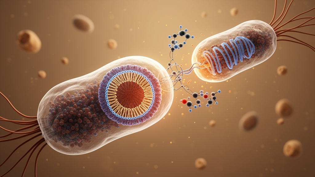
Microscopy and binding experiments indicated that the D configuration changed how the composite interacted with bacteria. Under illumination, treated bacteria showed collapsed membranes and leakage of intracellular contents, while nanoparticles were visibly attached to S. aureus cells. Fluorescein-labelled D-AuHNRs@CeO2 overlapped strongly with DNA staining in both bacterial species, whereas the L and mixed particles showed weaker overlap and remained largely outside the cells. Isothermal titration calorimetry gave the D composite lower dissociation constants for binding to the bacterial strains than the L and mixed configurations, consistent with stronger stereoselective association. D-AuHNRs@CeO2 also produced more heating near S. aureus after unbound particles were removed, suggesting that localization at the cell wall concentrated thermal energy. Quantification of nucleic acid and protein release showed increased leakage from both bacterial species, with especially high values after near-infrared irradiation and D-AuHNRs@CeO2 treatment. Together, these observations provided a mechanistic account for the stronger activity of the D formulation: preferential association with bacteria combined with more concentrated photothermal heating.
In mammalian cells, the D composite also performed better at reducing oxidative stress and promoting repair-related responses. After L929 fibroblasts were exposed to 2 millimolar hydrogen peroxide, DCFH-DA fluorescence and flow cytometry showed markedly lower intracellular reactive oxygen species after treatment with AuHNRs@CeO2; the D formulation produced the weakest signal. Scratch assays found greater migration by both L929 fibroblasts and human umbilical vein endothelial cells after 24 hours with D-AuHNRs@CeO2 than in the stressed controls. The D composite also increased messenger RNA expression of the antioxidant enzyme CAT and the repair-related factor TGF-β1 in L929 cells, and increased MMP1 expression in endothelial cells. These measurements showed effects on cultured-cell behaviour and gene expression under experimentally induced oxidative stress, rather than direct evidence of faster healing in patients. The investigators selected 100 micrograms per millilitre as the working concentration after hemolysis and cell-viability testing indicated that methoxypolyethylene glycol modification improved compatibility with living cells.
Cellular imaging suggested that oxidative stress influenced how the particles were taken up. Under stressed conditions, fluorescence from D-AuHNRs@CeO2 strongly colocalized with DAPI-stained cellular regions, while the L and mixed materials showed much less internalization; under normal culture conditions, signals from all three were concentrated around the cell periphery. In lipopolysaccharide-stimulated RAW264.7 macrophages, the D composite reduced expression of the M1-associated markers iNOS and IL-6 and increased the M2-associated markers CD206 and Arg-1. The authors interpreted these findings as evidence that oxidative-stress-dependent uptake enabled intracellular antioxidant activity and shifted macrophages toward a more pro-repair phenotype. The experiments were performed in cultured cell lines and did not establish how the same behavior would occur in an infected human wound. Nevertheless, the cellular results linked three measured functions: uptake of the D formulation, reduced oxidative stress and altered marker expression associated with macrophage polarization.
The strongest preclinical outcome came from infected full-thickness wounds in female BALB/c mice aged 6 to 8 weeks. Each 8-millimetre dorsal wound was inoculated with 0.1 millilitres of S. aureus at 10^7 colony-forming units per millilitre, and animals were randomly assigned to control, near-infrared, D-, L- or mixed-composite groups, with or without light. Treatments were applied on days 1 and 3, and wounds were monitored through day 12. D-AuHNRs@CeO2 plus near-infrared irradiation produced the fastest closure and the lowest bacterial counts, outperforming the non-irradiated groups and the corresponding L and mixed formulations. Colony counts were obtained from wound sites on days 3, 6, 9 and 12. A separate E. coli wound model, which included levofloxacin hydrochloride as a positive control, produced a comparable antibacterial ranking, with the D composite plus light showing the strongest effect among the tested nanoplatform groups. These results extended the antibacterial findings from culture assays to two bacterial infection models, while remaining limited to laboratory animals.
Histological examination was consistent with improved repair rather than simple suppression of infection. Control wounds retained heavy inflammatory infiltration, epidermal injury and loose connective tissue, whereas D-AuHNRs@CeO2 plus light showed a flatter wound surface, fewer inflammatory cells, nascent granulation tissue, new vessels, hair follicles and sebaceous glands. Masson’s trichrome staining showed that, by day 12, the same group had densely organized collagen fibers and a nearly continuous, thick epidermis with high keratinocyte density. Body weight changed only slightly across groups, and examinations of the heart, liver, spleen, lungs and kidneys reportedly showed no significant lesions. These observations support biocompatibility within the reported mouse experiments, but the material remains a preclinical experimental platform: the study did not test human wounds, and its findings do not demonstrate clinical benefit or establish whether the treatment would perform similarly in people.
Subject of Research: Preclinical testing of a chiral gold nanorod–cerium oxide nanoplatform for antibacterial and antioxidant wound healing.
Article Title: Synergistic photothermal and antioxidant effects of chiral gold helical nanorods @CeO 2 nanomaterials for accelerated infected wound healing
Article References: Xu, J., Li, Q., Kan, W., Liu, W., Liu, C., Liu, Q., Gao, J., Yan, K., Huang, C., & Wang, J. (2026). Synergistic photothermal and antioxidant effects of chiral gold helical nanorods @CeO2 nanomaterials for accelerated infected wound healing. Materials Today Bio, 41, Article 103754. https://doi.org/10.1016/j.mtbio.2026.103754
Image Credits: AI Generated
DOI: 10.1016/j.mtbio.2026.103754
Keywords: chiral nanomaterials, gold nanorods, cerium oxide, photothermal therapy, infected wounds, antibacterial materials, reactive oxygen species, tissue regeneration, macrophage polarization, mouse model, Synergistic, photothermal















 English (US) ·
 French (CA) ·