

 1 month ago
71
1 month ago
71
PROTECT YOUR DNA WITH QUANTUM TECHNOLOGY
Orgo-Life the new way to the future Advertising by AdpathwayIn a finding that reads like forensic chemistry applied to one of medicine’s most devastating infections, researchers have extracted and analyzed the metabolic fingerprints left in human brain tissue by fatal tuberculous meningitis, revealing that the tuberculosis bacterium appears to strip the brain of an extraordinary range of its chemical building blocks, from cholesterol and protective fatty acids to the very sugars needed to keep neurons functioning.
Tuberculous meningitis, or TBM, is the deadliest form of tuberculosis that spreads beyond the lungs. Caused by Mycobacterium tuberculosis, the same pathogen responsible for pulmonary TB, the disease inflames the delicate membranes surrounding the brain and spinal cord and produces distinctive immune clusters called granulomas in the brainstem and subarachnoid space. Even with the best available drug regimens, many patients die or are left with permanent neurological damage, and diagnosis remains notoriously difficult because the bacterium is present at extremely low levels in the cerebrospinal fluid, the clinical gold standard sample. A team of South African and Dutch researchers has now taken an unusual route to understanding what the pathogen does inside the brain itself: they studied tissue taken from patients who died of the disease, some archived nearly five decades ago.
The study, led by Abisola R. Isaiah of North-West University in Potchefstroom together with colleagues at Stellenbosch University and Amsterdam University Medical Center, drew on the archives of the neuropathology unit at Tygerberg Hospital in Cape Town. From collections spanning 1975 to 2012, the team assembled 41 formalin-fixed, paraffin-embedded, or FFPE, brain tissue samples from individual patients who died of TBM, along with 36 tissue blocks from six control patients who died of non-infectious causes. FFPE tissue is the workhorse of pathology departments worldwide, stored at room temperature for decades, but it has long been considered a hostile substrate for metabolomics, the comprehensive study of small molecules in biological systems. Fixation and paraffin embedding wash out some polar compounds and chemically alter others, which is why almost no metabolomics studies have attempted this material. Building on a standardized extraction protocol the group published previously, the researchers deparaffinized the tissue with xylene, melted the wax, homogenized roughly 20-milligram samples, and extracted metabolites in both polar and apolar fractions before chemically derivatizing them for analysis.
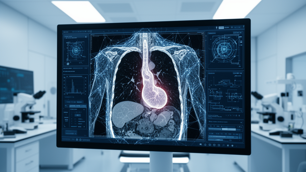
The analytical centerpiece was two-dimensional gas chromatography coupled to time-of-flight mass spectrometry, abbreviated GC×GC-TOFMS, a technique that separates compounds along two chromatographic dimensions and then identifies them by mass spectral fragmentation. This platform was chosen deliberately over proton nuclear magnetic resonance spectroscopy because it offers far greater sensitivity, an essential quality when working with milligram-scale tissue slices. For the TBM samples, the team went a step further in precision: a pathologist marked well-formed granulomas on stained slides, and an MMI CellCut laser microdissection system excised exactly those granulomatous regions from 20-micron-thick tissue sections, allowing the researchers to compare the immune clusters themselves with the surrounding brain tissue. Quality control samples pooled from control tissue were run at the beginning, middle, and end of each of eight analytical batches, and no instrumental drift was detected across the runs.
The statistical analysis revealed a striking separation between diseased and control tissue. In an unsupervised principal component analysis, the TBM and non-TBM groups clustered apart naturally, with the first two principal components together explaining just over 40 percent of the variance. A supervised partial least squares-discriminant analysis performed even more impressively, with cross-validated R-squared and Q-squared values of 0.89 and 0.87, prediction accuracy of 97.8 percent, and permutation testing confirming the model was not overfitting. Out of 454 metabolites detected, 294 drove this separation, and 41 metabolites passed stringent significance thresholds combining multivariate importance scores above 1.0, fold changes greater than 2.0, false-discovery-rate-corrected p-values below 0.05, and effect sizes exceeding 0.8.
What the team found was a portrait of profound chemical depletion. Ten metabolites, including arachidonic acid, D-chiro-inositol, pipecolic acid, pregnenolone, and L-pyroglutamic acid, fell below the detection limit entirely in every TBM case. The most dramatic single observation concerned cholesterol: control brain tissue contained about 96.86 milligrams per gram, while TBM tissue held roughly a hundredfold less, at 0.58 milligrams per gram. Because the brain is the most cholesterol-rich organ in the body and M. tuberculosis is known to harvest host cholesterol for its own energy and persistence, the authors suggest the bacterium may be consuming nearly all of the brain’s cholesterol supply in terminal disease.
The hydrocarbons told a similarly compelling story. Alkanes and alkenes, compounds that occur naturally in brain myelin, were either sharply depleted or absent in TBM samples. The researchers propose two non-exclusive explanations: altered host lipid metabolism and tissue injury, or direct scavenging by the bacterium, which has been shown to catabolize these molecules as a carbon source. Consistent with the latter idea, comparisons between the microdissected granuloma regions and surrounding tissue identified six differentially abundant metabolites, all of them hydrocarbons, although the effect sizes in that granuloma-specific comparison were small.
The lipid losses extended across the board. Oleic acid, essential for myelin synthesis but known to acidify the local environment in ways that favor mycobacterial growth, was depleted and negatively correlated with final disease staging, meaning the more severe the disease, the less of it remained. Arachidonic acid, a keystone of the inflammatory response released by phospholipase A2, and linoleic acid, its membrane-protective precursor, were both significantly reduced. Pregnenolone, a neurosteroid that suppresses overactive macrophages during inflammation, vanished below detection limits, as did trans-vaccenic acid, a diet-derived fatty acid that mycobacteria are known to consume as nutrition.
Perhaps most intriguing for future drug development is what happened to the lysine degradation pathway. Metabolites connected to this route, including glutaric acid, pipecolic acid, piperidine, 2-oxoadipic acid, and 2-oxoglutarate, were significantly altered, and the authors highlight lysine catabolism as a plausible alternative energy source enabling the pathogen to persist in a dormant state within the oxygen-starved environment of a granuloma. M. tuberculosis is famous for its ability to survive hypoxia without replicating, and the team speculates that in the severely hypoxic granuloma the bacterium may actually scavenge oxygen from host fatty acids, transferring it to hydrocarbons to fuel replication. Prior work has suggested that inhibiting the aspartate pathway, from which lysine is biosynthesized, could help eradicate persistent infection, making the new brain-tissue evidence a potentially significant corroboration.
The sugar and alcohol story carries direct clinical resonance. Glucose was detectable at roughly 10 milligrams per gram in control tissue but absent entirely in TBM samples. Myo-inositol and D-chiro-inositol, sugar alcohols with neuroprotective and insulin-mimetic properties that the bacterium reportedly incorporates into its cell wall, were severely depleted, and the authors connect this loss to the confusion and delirium seen in advanced patients. Meanwhile, several alcohols with antimicrobial properties, including 2-phenylethanol and hexanol, dropped dramatically. Notably, 2-phenylethanol showed positive associations with brainstem infarcts and with granulomas in the infratentorial regions and cranial nerve roots in the team’s network analysis, hinting it might serve as a marker of bacterial presence.
The researchers are careful about the limitations. The control tissue was not entirely healthy, age distributions differed between groups, and samples came from heterogeneous brain regions. FFPE degradation over decades remains a confound, all compound identifications are putative at Metabolomics Standards Initiative level 2, and the small number of control patients means statistical analyses relied on tissue blocks rather than patient counts. Correlations between metabolites and clinical variables, such as fumaric acid’s positive relationship with bacillary load, were statistically weak and framed as hypothesis-generating rather than confirmatory.
Even with those caveats, the study represents a first. To the authors’ knowledge, no prior metabolomics analysis has been performed on brain tissue from TBM cases, and the results dovetail with earlier findings in urine and cerebrospinal fluid from the same research group and others, suggesting that the metabolic havoc wreaked by the bacterium is consistent across sample types and can be traced from the organ itself outward to accessible biofluids. If validated, the depleted or absent metabolites, particularly those vanishing entirely in terminal disease, could anchor new biomarker panels capable of distinguishing TBM from other forms of meningitis at earlier, more treatable stages. Just as importantly, the work demonstrates that pathology archives, those vast repositories of wax-embedded tissue sitting in hospital basements around the world, hold recoverable chemical information that modern analytical instruments are only now learning to read.
Subject of Research: Metabolic characterization of tuberculous meningitis in postmortem FFPE human brain tissue using untargeted GC×GC-TOFMS metabolomics
Subject of Research: Biology
Article Title: Metabolic insights into the pathophysiology of tuberculous meningitis in FFPE postmortem human brain tissue
Article References: Isaiah, A. R., Loots, D. T., Williams, A. A., Zaharie, S. D., van Furth, A. M. T., van der Kuip, M., & Mason, S. (2026). Metabolic insights into the pathophysiology of tuberculous meningitis in FFPE postmortem human brain tissue. Metabolomics, 22(4), Article 138. https://doi.org/10.1007/s11306-026-02511-8
Image Credits: AI Generated
DOI: 10.1007/s11306-026-02511-8
Keywords: tuberculous meningitis, Mycobacterium tuberculosis, metabolomics, FFPE brain tissue, granuloma, GC×GC-TOFMS, cholesterol depletion, lysine degradation pathway, biomarkers, central nervous system tuberculosis
Cite Scienmag News
APA MLA Chicago
Cassandra Pierce. (September 6, 2026). Metabolic changes reveal how tuberculosis damages the postmortem human brain. Scienmag. https://scienmag.com/metabolic-changes-reveal-how-tuberculosis-damages-the-postmortem-human-brain/
Copy citation Download RIS
Tags: brain tissue analysis in tuberculosischallenges in diagnosing tuberculous meningitischemical alterations in tuberculous brain tissueeffects of TB on neuronal function and lipidseffects of tuberculosis on neuronal function and brain lipidsforensic chemistry in infectious disease researchforensic chemistry of infectious brain diseasesgranulgranuloma formation in tuberculous brain infectionhistorical brain tissue studies in infectious diseaseshistorical tissueimpact of Mycobacterium tuberculosis on brain chemistrylong-term neurological damage from tuberculosislow-level detection of Mycobacterium tuberculosis in cerebrospinal fluidmetabolic changes caused by tuberculosis infectionmetabolic changes in brain due to tuberculous meningitismetabolic depletion in infected brain tissueneurochemical damage caused by tuberculosis infectionneurological damage from tuberculous meningitispostmortem brain damage in tuberculosispostmortem brain tissue analysis in tuberculosisTuberculous meningitis metabolic fingerprint













 English (US) ·
  French (CA) ·