
 2 hours ago
1
2 hours ago
1
PROTECT YOUR DNA WITH QUANTUM TECHNOLOGY
Orgo-Life the new way to the future Advertising by Adpathway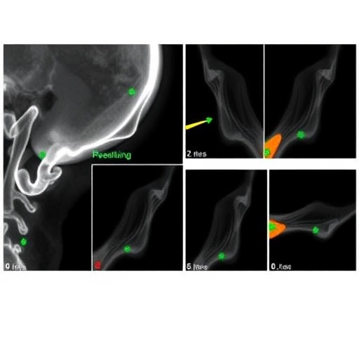
In a groundbreaking study, researchers have successfully applied advanced deep learning techniques to classify the morphological variations of the mandibular condyle as observed through panoramic radiographs. The mandibular condyle, a vital component in the temporomandibular joint, plays an essential role in mastication and overall jaw function. Any morphological discrepancies can lead to significant clinical implications, including pain, dysfunction, and even surgical interventions. This pioneering research paves the way for more effective diagnostic methodologies and has the potential to transform our approach to dental radiology.
The study, which appears in the esteemed Journal of Medical and Biological Engineering, introduces a sophisticated algorithm tailored to analyze panoramic radiographs for classifying different morphological types of the mandibular condyle. This technique seeks not only to enhance diagnostic accuracy but also to minimize human error, which can often arise during manual assessments. By using an automated system driven by deep learning, the research underscores the potential of artificial intelligence in revolutionizing traditional medical practices.
For a long time, clinicians have relied on their expertise and experience to interpret panoramic radiographs. However, variations in training and individual judgement can lead to inconsistencies in diagnoses. The researchers propose that a deep learning approach can standardize the assessment process, offering a scalable solution that delivers uniform outcomes across various clinical settings. By leveraging vast datasets of radiographic images, the algorithm learns to identify subtle differences in the morphology of the mandibular condyle with unprecedented precision.
In this recent study, the researchers adopted convolutional neural networks (CNNs), a class of deep learning models particularly suited for image processing tasks. These networks efficiently capture spatial hierarchies in images, making them ideal for the analysis of complex anatomical structures. After training on a diverse array of panoramic radiographs, the CNN was able to discern minute features that could signify different morphological variations of the mandibular condyle, including hyperplasia, atrophy, and various asymmetries.
Throughout the study, validation was conducted to measure the algorithm’s performance against traditional human interpretation. The results showed that the deep learning model not only matched but often exceeded the accuracy of seasoned practitioners. This finding bolsters the argument for incorporating artificial intelligence into diagnostic radiology, highlighting the potential to enhance patient care. Moreover, with the algorithm’s ability to continually learn and adapt, its performance is likely to improve as more data becomes available.
One of the critical aspects of this research is its recognition of the intricate variations that exist in the human anatomy. The mandibular condyle can exhibit a spectrum of shapes and sizes, influenced by genetic, developmental, and environmental factors. By categorizing these variations systematically, the study offers a framework that could help clinicians predict potential functional complications stemming from specific morphological traits. This predictive power could ultimately lead to tailored treatment plans for individual patients, improving their overall health outcomes.
In addition to practical applications in clinical dental practice, this research also opens avenues for further studies that could explore the underlying genetic and developmental mechanisms responsible for the observed morphological variations. By combining deep learning techniques with genetic data, future research might delve deeper into the etiology of these variations, potentially identifying biomarkers for predisposition to mandibular joint disorders. This holistic understanding could inform preventive strategies in dental care and orthodontics.
This innovative approach underscores the growing intersection of technology and healthcare, where artificial intelligence is not only augmenting human capabilities but also redefining how medical professionals approach diagnosis and treatment. The potential for deep learning applications extends beyond dentistry; it sets a precedent for other fields in medicine where image analysis is pivotal. The algorithm used in this study could inspire similar advancements in areas such as radiology, pathology, and orthopedics.
Furthermore, the implications of this research extend into the educational realm. As budding dentists and radiologists equip themselves with knowledge of deep learning principles, the integration of AI in their training could prepare them for a future where technology is indispensable to their practice. This shift not only enhances their skill set but also instills a sense of confidence in leveraging AI tools to support clinical decisions.
The journey to this breakthrough reflects a collaborative effort among various fields of expertise, including dental medicine, biomedical engineering, and computer science. Such interdisciplinary partnerships are crucial for driving innovation and addressing complex health issues. The convergence of these domains is essential in developing new technologies that are not only effective but also practical for everyday clinical use.
Overall, the application of deep learning in classifying mandibular condyle variations signifies a substantial leap toward the convergence of artificial intelligence and medical diagnostics. As we move into an era where such innovations become the norm, the research not only highlights the efficiency of technology but also reaffirms the importance of human oversight in interpreting the outcomes of AI-driven analyses. The combination of both elements promises to yield optimal diagnosis and treatment pathways for patient care in the years to come.
As the field of dental radiology continues to evolve, this research stands as a testament to the endless possibilities that await when we harness the power of technology alongside clinical expertise. The future of diagnosis is bright, and the implications for patient welfare are immeasurable, ensuring that advancements today will ripple through the healthcare sector for generations.
Subject of Research: Deep Learning Approaches for Classifying Morphological Variations of the Mandibular Condyle
Article Title: Classification of Morphological Variations of Mandibular Condyle in Panoramic Radiographs with a Deep Learning Approach
Article References:
Yuce, F., Öziç, M.Ü. & Buyuk, C. Classification of Morphological Variations of Mandibular Condyle in Panoramic Radiographs with a Deep Learning Approach.
J. Med. Biol. Eng. (2025). https://doi.org/10.1007/s40846-025-00962-3
Image Credits: AI Generated
DOI: 10.1007/s40846-025-00962-3
Keywords: Deep Learning, Machine Learning, Panoramic Radiographs, Morphological Variations, Mandibular Condyle, Artificial Intelligence, Dental Radiology, Convolutional Neural Networks.
Tags: advanced algorithms in healthcareArtificial Intelligence in Medicineautomated diagnostic techniquesclinical implications of jaw morphologydeep learning in dental radiologyinnovative approaches to dental diagnosticsmandibular condyle classificationmorphological variations of jawbonepanoramic radiograph analysisreducing human error in radiologystandardizing medical assessmentstemporomandibular joint disorders


 English (US) ·
  French (CA) ·