

 1 hour ago
8
PROTECT YOUR DNA WITH QUANTUM TECHNOLOGY
Orgo-Life the new way to the future Advertising by AdpathwayAdvanced ultrasound analysis utilizing two-dimensional speckle-tracking imaging can identify occult biventricular and left atrial cardiac deformation in patients diagnosed with liver cirrhosis well before conventional echocardiographic techniques show resting anomalies, according to a clinical investigation conducted by medical researchers at the Third Affiliated Hospital of Qiqihar Medical University. Cirrhotic cardiomyopathy, formally defined at the 2005 World Congress of Gastroenterology by impaired contractile reserve, electrophysiological irregularities, and diastolic dysfunction, often remains clinically undetectable during routine medical examinations because a pronounced hyperdynamic circulatory state masks latent myocardial injury.
To evaluate these subclinical myocardial changes across advancing stages of liver failure, investigators initially recruited ninety-six consecutive individuals diagnosed with liver cirrhosis who presented to the Department of Gastroenterology and Infectious Diseases between July 2024 and January 2025. Because direct ethanol consumption induces known myocardial toxicity independent of chronic liver failure, all individuals presenting with alcoholic cirrhosis were deliberately excluded to maintain clinical homogeneity within the cohort. Strict exclusionary standards further eliminated participants presenting with primary cardiac diseases, including coronary artery disease, valvular heart conditions, known cardiomyopathies, atrial fibrillation, and congenital structural abnormalities, as well as those with chronic systemic conditions such as arterial hypertension, diabetes mellitus, thyroid disturbances, chronic renal impairment, and chronic respiratory disorders. Following the exclusion of secondary pathologies and the removal of seven individuals whose acoustic windows prevented adequate echocardiographic tracking, eighty-two patients comprising sixty-two men and twenty women with a mean age of 58.34 years entered the observation group. Thirty healthy individuals frequency-matched by age, sex, height, and weight served as control subjects after two candidates were removed for suboptimal acoustic tracking.
The eighty-two enrolled patients with liver cirrhosis were classified according to the standardized Child-Pugh grading rubric, which calculates clinical severity based on serum albumin levels, total bilirubin concentrations, prothrombin time prolongation, and the physical presence of ascites or hepatic encephalopathy. This staging assigned thirty participants to Class A representing early disease, twenty-nine to Class B indicating intermediate impairment, and twenty-three to Class C reflecting decompensated hepatic failure. Basic demographic markers, including chronological age, gender distribution, body mass index, standing height, body weight, resting blood pressure, and heart rate, demonstrated no statistically significant differences between the healthy control group and the three cirrhotic subgroups, ensuring that baseline hemodynamic discrepancies did not account for observed variations. Laboratory evaluations confirmed expected biochemical deteriorations across the clinical spectrum, with serum albumin decreasing systematically across groups while prothrombin time and total bilirubin exhibited marked, statistically significant elevations as patients advanced from compensated status to decompensated Class B and Class C stages.
All transthoracic examinations were completed using a Philips EPIQ 7 ultrasound system fitted with an S5-1 phased-array transducer operating at frequencies between one and five megahertz, with subjects positioned in the standard left lateral decubitus orientation during end-expiratory breath-holding. Digital loops of at least three consecutive cardiac cycles were archived in native DICOM format at acquisition rates exceeding sixty frames per second from standard apical four-chamber, three-chamber, and two-chamber views. A blinded sonographer analyzed dynamic loops using dedicated automated strain algorithms designed specifically for the left ventricle, right ventricle, and left atrium, successfully achieving analytical feasibility in 92.5 percent of attempted cases across the entire study cohort. Whenever automated border tracking exhibited drift or border ambiguity in segments displaying lower speckle quality, manual endocardial adjustments were applied, a step required in thirty-three percent of cases. Repeat evaluations on a random subset of twenty-five re-examined patients confirmed high intra-observer and inter-observer reproducibility through rigorous Bland-Altman consistency analyses.
Standard transthoracic echocardiographic examinations demonstrated notable limitations when screening for early myocardial damage, failing to differentiate between cirrhotic patients and healthy controls on multiple standard functional metrics. Left ventricular ejection fraction, interventricular septal thickness at end-diastole, left ventricular posterior wall thickness at end-diastole, and the pulsed-wave Doppler ratio of early to late peak diastolic mitral inflow velocities revealed no statistically significant differences across any study cohorts. This stability in resting ejection fraction highlights how the hyperdynamic circulation characteristic of liver cirrhosis maintains normal or even supranormal volumetric pumping performance despite underlying cellular injury. Standard signs of diastolic impairment and dimensional remodeling emerged almost exclusively in advanced disease stages, where the tissue Doppler early diastolic velocity at the septal mitral annulus dropped significantly and the ratio of early mitral inflow to annular tissue velocity increased markedly in Classes B and C, accompanied by chamber dilation in Class C.
Conventional assessments of right ventricular geometry and systolic mechanics mirrored this late-stage pattern of structural and functional deterioration, showing normal values throughout early cirrhosis and sudden declines in decompensated disease. Standard right ventricular performance indices, including tricuspid annular plane systolic excursion, tissue Doppler peak systolic velocity at the tricuspid annulus, and right ventricular fractional area change, remained statistically preserved across Class A and Class B cohorts before exhibiting steep, statistically significant drops among Class C patients. Structural chamber dimensions displayed reciprocal expansions under standard acoustic calipers, with right ventricular basal diameter, right atrial transverse diameter, and right atrial longitudinal diameter showing significant increases exclusively within the decompensated Class C subgroup. These late alterations demonstrate that routine dimensional metrics and conventional annular excursion indices cannot reliably register the insidious remodeling that takes place before liver cirrhosis progresses into severe hepatic decompensation.
In sharp contrast to traditional acoustic measurements, two-dimensional speckle-tracking imaging unmasked significant mechanical dysfunction during early, compensated hepatic disease. Left ventricular global longitudinal strain and right ventricular free wall longitudinal strain were significantly diminished across all cirrhotic subgroups relative to healthy controls, displaying a progressive downward trajectory from Class A through Class C. Early myocardial degradation was particularly evident on the right side of the heart, where right ventricular free wall strain exhibited significant reduction in Class A patients, whereas right ventricular global longitudinal strain, which incorporates the interventricular septum, showed statistically significant reductions only after patients advanced into Classes B and C. Spearman rank correlation testing documented significant negative correlations between Child-Pugh severity scores and left ventricular global longitudinal strain, right ventricular free wall longitudinal strain, and right ventricular global longitudinal strain, demonstrating a direct numerical link between liver failure severity and worsening ventricular contractility.
The pronounced vulnerability of the right ventricular free wall during early disease reflects distinct regional mechanical pressures and anatomical architecture. On a cellular level, cirrhotic cardiomyopathy stems from several interacting pathological mechanisms, including the downregulation of beta-adrenergic receptor density, impaired cyclic adenosine monophosphate production, altered cardiomyocyte membrane fluidity, and systemic accumulation of endogenous cardiodepressant substances such as nitric oxide and endocannabinoids. While occasional published studies have observed elevated global longitudinal strain in early cirrhosis due to hyperdynamic hypercontractility, the predominance of advanced cases and software variations between ultrasound equipment vendors explain divergences across medical literature, reinforcing the progressive contractile decline seen across structured clinical tiers.
Speckle-tracking interrogation of left atrial strain unveiled a distinct tripartite mechanical response that dynamically reflects progressive left ventricular diastolic stiffness across advancing cirrhosis stages. Left atrial conduit strain, which measures passive blood transit into the ventricle during early diastole, declined steadily and significantly from healthy controls to Class A, Class B, and Class C cohorts, demonstrating that left ventricular suction capability is impaired even in compensated cirrhosis. In contrast, left atrial reservoir strain and contractile booster-pump strain displayed a clear biphasic compensatory pattern, rising significantly above healthy control values in Class A and Class B patients to sustain ventricular filling against deteriorating conduit mechanics. However, when patients progressed into decompensated Class C disease, this compensatory reserve was completely exhausted, resulting in severe drops in both reservoir and contractile strain values compared to healthy individuals and earlier cirrhosis subgroups, directly signaling mechanical atrial failure under unrelenting pressure overload.
The clinical investigators noted several methodological limitations, pointing out that the cross-sectional design prevents the determination of longitudinal causal pathways and limits prospective prognostic conclusions regarding cardiovascular events or mortality. Furthermore, the decompensated Class C cohort comprised twenty-three participants whose Child-Pugh scores clustered between 11 and 12, omitting critically ill patients scoring between 13 and 16 and thereby constraining the generalizability of findings in terminal liver disease. Nevertheless, these findings demonstrate that two-dimensional speckle-tracking imaging provides an objective, angle-independent diagnostic modality capable of exposing subclinical biventricular and atrial strain impairments during early cirrhosis, offering valuable hemodynamic insights to guide timely clinical decision-making prior to major surgical interventions such as liver transplantation.
Subject of Research: Two-dimensional speckle-tracking echocardiography for early cardiac dysfunction assessment in liver cirrhosis
Article Title: Two-dimensional speckle-tracking imaging for assessing cardiac dysfunction in liver cirrhosis: correlation with Child-Pugh score
Article References: Ma, J., Ding, Y., Shao, H., Zhang, B., & Jiang, C. (2026). Two-dimensional speckle-tracking imaging for assessing cardiac dysfunction in liver cirrhosis: correlation with Child-Pugh score. Cardiovascular Ultrasound, 24(1), Article 29. https://doi.org/10.1186/s12947-026-00384-4
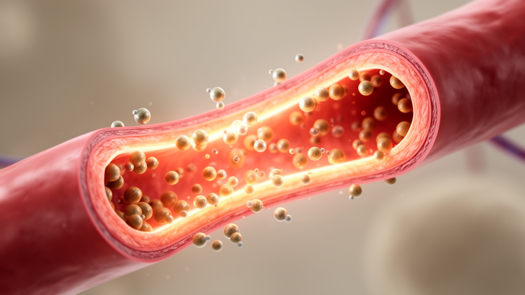
Image Credits: AI Generated
DOI: 10.1186/s12947-026-00384-4
Keywords: liver cirrhosis, two-dimensional speckle tracking imaging, cirrhotic cardiomyopathy, left ventricular global longitudinal strain, right ventricular free wall longitudinal strain, right ventricular global longitudinal strain, left atrial conduit strain, left atrial reservoir strain, left atrial contractile strain, Child-Pugh classification, conventional echocardiography, diastolic dysfunction, myocardial deformation, tissue Doppler imaging, tricuspid annular plane systolic excursion, subclinical cardiac dysfunction, liver transplantation assessment
















 English (US) ·
 French (CA) ·