

 5 hours ago
7
PROTECT YOUR DNA WITH QUANTUM TECHNOLOGY
Orgo-Life the new way to the future Advertising by AdpathwayResearchers have established a comprehensive and scalable workflow for applying volume electron microscopy to human cortical organoids, marking a significant advancement in the ability to analyze neural circuits at the ultrastructural level. Published in Cellular and Molecular Neurobiology, this original research demonstrates for the first time the feasibility of micro-connectomic reconstruction in human cortical organoids at nanometer resolution. The study addresses a critical limitation in current neuroscience, where the application of volume electron microscopy to human brain organoids has remained limited, particularly for connectomic studies that require detailed mapping of synaptic connections.
Their approach combines correlative light and electron microscopy with large-area scanning electron microscopy mosaic imaging. This is followed by focused ion beam–scanning electron microscopy and validated by transmission electron microscopy. By systematically comparing two embedding protocols currently in use, the researchers identified specific technical advantages and limitations that influence the quality of ultrastructural data obtained from these complex biological samples.
A central finding of the study involves the comparison of embedding methods, specifically the DeFelipe and Fairén method versus the Deerinck protocol. The authors demonstrated that the DeFelipe and Fairén method provides optimal compatibility with toluidine blue–stained semithin sectioning. This compatibility enables reliable synapse segmentation and neurite tracing, which are essential for accurate connectome reconstruction. In contrast, while the Deerinck protocol offers enhanced membrane contrast, it limits the visualization of postsynaptic densities, thereby restricting its utility for certain types of synaptic analysis.
Using focused ion beam–scanning electron microscopy imaging of peripheral, neuropil-like regions of the cortical organoids, the researchers achieved accurate three-dimensional reconstruction of synapses, neurites, and intracellular organelles. This high-resolution imaging allowed for a quantitative assessment of synaptic apposition surfaces, neurite trajectories, and organelle distribution across defined cellular compartments.
The methodological framework developed in this study expands the applicability of volume electron microscopy to organoid systems. By establishing a pipeline that moves from sample preparation and resin embedding to semithin and ultrathin sectioning, the team created a scalable process for connectome reconstruction. Large-area scanning electron microscopy mosaic imaging was used to identify regions of interest for targeted focused ion beam–scanning electron microscopy acquisition, ensuring that the most relevant areas were analyzed in high detail.
The implications of this work extend beyond methodological development, providing a robust foundation for future studies of human brain development, disease modeling, and therapeutic evaluation at the synaptic and subcellular level.
The study highlights the potential of organoid systems as a platform for high-resolution connectomic analysis.
Subject of Research: Methodological development for volume electron microscopy and connectome reconstruction in human cortical organoids
Article Title: Volume Electron Microscopy of Cortical Organoids: Methods for Region Identification, Connectome Reconstruction, and Organelle Segmentation
Article References: Dallere, S., Mattioni, A., Turégano-López, M., Blazquez-Llorca, L., Merchan-Perez, A., Schellino, R., Vercelli, A., DeFelipe, J., & Boido, M. (2026). Volume Electron Microscopy of Cortical Organoids: Methods for Region Identification, Connectome Reconstruction, and Organelle Segmentation. Cellular and Molecular Neurobiology. https://doi.org/10.1007/s10571-026-01814-7
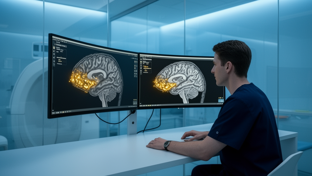
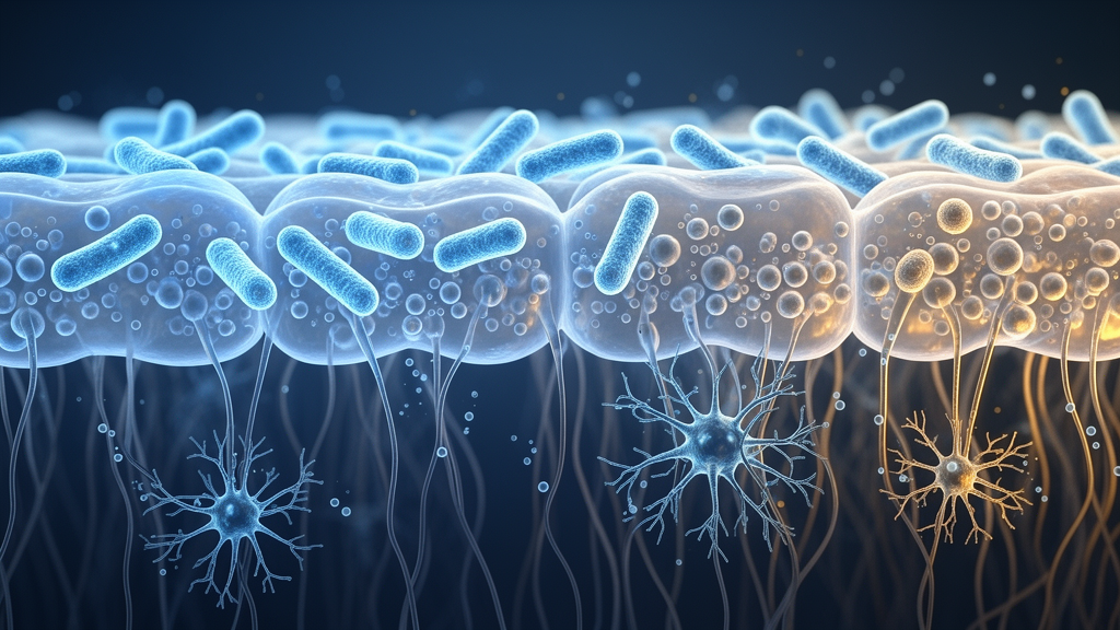
Image Credits: AI Generated
DOI: 10.1007/s10571-026-01814-7
Keywords: volume electron microscopy, cortical organoids, connectome reconstruction, FIB-SEM, synaptic analysis, neuroscience, ultrastructure, human brain development, Volume, Electron, Microscopy, Cortical












 English (US) ·
 French (CA) ·