

 3 days ago
9
PROTECT YOUR DNA WITH QUANTUM TECHNOLOGY
Orgo-Life the new way to the future Advertising by AdpathwayIn an extensive review published in Frontiers in Cell and Developmental Biology, an international research team led by investigators from the Schepens Eye Research Institute of Massachusetts Eye and Ear and Harvard Medical School synthesized current mechanistic evidence on Rho-associated coiled-coil containing protein kinases 1 and 2, commonly designated as ROCK1 and ROCK2. These downstream effectors of RhoA occupy a pivotal position at the biochemical and mechanical crossroads where tissue scarring and neural decline converge. Although fibrosis has traditionally been defined by excessive extracellular matrix deposition, progressive tissue stiffening, and organ architectural disruption, neurodegenerative conditions are primarily characterized by synaptic failure, neuronal dysfunction, impaired regenerative capacity, and progressive cell loss. Despite these categorical distinctions, the authors show that both disease categories share fundamental hallmarks of maladaptive cellular reprogramming, persistent chronic stress, impaired homeostatic resilience, and progressive structural disorganization. These shared pathological features are especially pronounced within ocular and central nervous system compartments, where cellular health depends closely on tightly regulated physical forces, tissue stiffness, and intact biomechanical architecture.
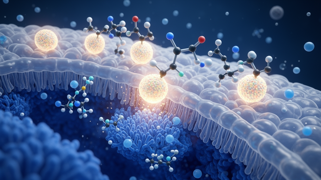
Under normal physiological conditions, ROCK-mediated mechanotransduction is not inherently destructive or maladaptive. Controlled RhoA-ROCK activity functions as a dynamic physical sensory system, enabling cells to detect subtle changes in matrix rigidity, cell geometry, extracellular adhesion, and externally applied mechanical load. Through the direct phosphorylation of myosin regulatory light chain and the concurrent inhibitory phosphorylation of myosin phosphatase, ROCK increases actomyosin contractility, while downstream ROCK and LIM kinase signaling simultaneously prevents cofilin-mediated actin depolymerization to stabilize actin filaments. In healthy ocular and neural environments, these precise biomechanical processes maintain cortical tension, regulate cell shape, stabilize intercellular junctions and cell-matrix adhesions, preserve endothelial and epithelial barrier integrity, modulate smooth-muscle tone, and sustain architectural resilience against mechanical stress. Following acute trauma, transient ROCK activation drives productive wound healing by coordinating directional cell migration, epithelial closure, provisional matrix assembly, and tissue contraction, as demonstrated in cutaneous and corneal repair models. Once tissue continuity is restored, declining inflammatory signals, reduced mechanical strain, and the deactivation or clearance of temporary myofibroblasts normally resolve pathway activity, preventing reparative remodeling from evolving into persistent tissue fibrosis.
When these normal termination pathways fail, unresolved inflammation, elevated transforming growth factor-beta signaling, matrix stiffening, and excessive cell-generated mechanical tension combine to lock cells into chronic contractile loops. Studies of peripheral fibrotic disease across pulmonary, hepatic, cardiac, and dermal tissues demonstrate that ROCK-dependent actomyosin contractility continually reinforces myofibroblast differentiation, sustained matrix production, and bidirectional fibrogenic crosstalk with inflammatory cells. Investigations using human scleroderma fibroblasts and experimental pulmonary fibrosis models have confirmed that both ROCK1 and ROCK2 participate in these reinforcing feedback loops. However, the review emphasizes that peripheral fibrosis models provide comparative context rather than proof of identical biology within the central nervous system or the eye. While physical forces such as hydrostatic pressure, tensile stretch, fluid shear, and matrix stiffness regulate trabecular meshwork cells, Schlemm canal endothelia, Müller glia, and optic nerve head glia in the eye, as well as cerebral glia and neuronal networks in the brain, neurodegenerative pathology cannot simply be equated with peripheral fibrosis. Therapeutic evaluation therefore demands careful dissection of cell types, injury stages, and anatomical locations.
A critical bridge linking mechanical forces to enduring pathological changes is the interaction between ROCK signaling and force-sensitive transcriptional regulators, specifically YAP/TAZ and MRTF/SRF. Increased matrix rigidity, cellular spreading, and cytoskeletal tension promote the nuclear translocation of Yes-associated protein and transcriptional coactivator with PDZ-binding motif, partly via direct physical force transmission through nuclear pores. In parallel, myocardin-related transcription factors remain sequestered in the cytoplasm by binding globular G-actin; when RhoA-ROCK signaling accelerates actin polymerization into filamentous F-actin, the depletion of free G-actin allows MRTF to translocate into the nucleus and activate serum response factor-dependent gene transcription. Experimental studies in human pterygium fibroblasts and scleral fibroblasts exposed to mechanical strain or transforming growth factor-beta demonstrate that ROCK inhibition directly blocks MRTF-A nuclear translocation and suppresses downstream fibrogenic gene expression programs. However, evidence directly connecting ROCK to YAP/TAZ in ocular tissues remains less extensive than general mechanobiology observations, and the authors caution that suppressing these transcriptional modules indiscriminately could harm neural or reparative glial cells where YAP/TAZ or MRTF/SRF activity supports structural repair and regeneration.
Ocular fibrocontractile conditions highlight the complex translational challenges of targeting this pathway. In proliferative vitreoretinopathy, a sight-threatening complication occurring after retinal detachment and repair, retinal pigment epithelial cells undergo phenotypic transdifferentiation into traction-generating contractile cells that distort the retinal architecture. Experimental studies show that combining ROCK and transforming growth factor-beta pathway inhibition suppresses alpha-smooth muscle actin and fibronectin expression while maintaining epithelial tight-junction markers such as ZO-1, and specific ROCK-inhibiting formulations attenuate experimental proliferative vitreoretinopathy. Beyond retinal membranes, pharmacological ROCK modulation demonstrates tissue-specific benefits in other ocular compartments. In glucocorticoid-induced ocular hypertension models, the clinically established inhibitor netarsudil lowers trabecular meshwork stiffness, increases aqueous outflow facility, and reduces histologic scarring markers. Similarly, in choroidal neovascularization-associated subretinal fibrosis models, the pan-ROCK inhibitor fasudil and the ROCK2-selective inhibitor belumosudil reduce fibrotic scarring and phosphorylated MYPT1 levels. Nevertheless, because experimental results from one compartment cannot guarantee clinical efficacy across another, translating these findings into validated treatments requires direct posterior-segment exposure and disease-specific confirmation.
The retina provides a uniquely informative biological testing ground because neuronal destruction and fibrocontractile scarring frequently develop within the exact same anatomical microenvironment. Following retinal detachment, mechanical detachment causes immediate photoreceptor stress and synaptic dissociation, often alongside the aberrant cellular proliferation that produces tractional membranes. Experimental investigations demonstrate that treatment with fasudil stabilizes rod photoreceptor synaptic terminals and reduces structural damage in the outer plexiform layer after experimental detachment. In complementary optic nerve and retinal injury studies, topical administration of the inhibitor AR-13324 increased retinal ganglion cell survival and promoted axon regeneration, while intravitreal netarsudil provided substantial axonal protection in tumor necrosis factor-induced neurodegeneration models. These experimental findings indicate that carefully timed ROCK pathway inhibition might simultaneously counteract tractional fibrocontractile scarring while shielding delicate retinal neurons from secondary degeneration. However, the authors underscore that despite promising experimental data, clinical studies have not yet demonstrated simultaneous antifibrotic and neuroprotective efficacy within human patients receiving a single therapeutic intervention.
In the brain and spinal cord, RhoA-ROCK signaling participates directly in regenerative failure following trauma and during chronic neurodegenerative progression. Following central nervous system damage, myelin-associated inhibitory molecules—including Nogo-A, myelin-associated glycoprotein, and oligodendrocyte myelin glycoprotein—bind neuronal surface receptors and activate intracellular RhoA-ROCK signaling cascades. This activation stimulates LIM kinase and myosin light chain phosphorylation, triggering rapid actomyosin contraction, growth-cone collapse, and persistent axonal retraction. Pharmacological ROCK inhibition counteracts this inhibitory cascade in experimental models by reducing cytoskeletal tension and allowing microtubules to advance into extending growth cones. Concurrently, ROCK regulates synaptic plasticity by controlling actin turnover within dendritic spines, where excessive pathway activity causes spine shrinkage, synaptic destabilization, and impaired long-term potentiation. Moreover, elevated ROCK activity promotes amoeboid, pro-inflammatory microglial phenotypes that release reactive oxygen species and inflammatory cytokines, whereas pharmacological inhibition reduces microglial inflammatory mediator production and alters motility. Consequently, experimental neural protection from ROCK inhibition reflects a combination of neuron-intrinsic regeneration, synaptic spine stabilization, and suppression of harmful neuroinflammation.
Crucially, the review warns against viewing central nervous system injury-associated scarring through an oversimplified antifibrotic lens. Traumatic neural scars contain biologically distinct components that execute divergent functions during tissue repair. For example, experimental spinal cord injury models indicate that while moderately attenuating pericyte-derived stromal fibrotic scars can promote descending axonal regeneration and improve functional sensorimotor recovery, preventing or eliminating astrocytic scar formation severely disrupts tissue repair and fails to support spontaneous axon regrowth. Astrocytes form protective borders that isolate damaged areas and support regenerating fibers, a process heavily reliant on mechanosensitive signaling. Indeed, experimental studies show that astrocytic YAP activation promotes organized glial border assembly, axonal regrowth, and functional recovery after spinal cord injury, whereas astrocytic YAP deletion causes severe architectural disintegration. Indiscriminately abolishing scar-forming programs or downstream transcriptional networks could therefore undermine essential neuroprotective barriers. Determining whether stromal and immune ROCK activation can be selectively inhibited without compromising beneficial astrocytic scar formation remains an unresolved priority in neurotrauma research.
A critical consideration highlighted by the authors is the biological specialization of ROCK1 versus ROCK2 across distinct cell states. Both kinase isoforms share structural homology and core phosphorylation targets, yet single-cell transcriptomic analyses demonstrate that they regulate overlapping but non-identical coding and noncoding gene networks. For instance, ROCK1 activity has been specifically implicated in diabetic endothelial and retinal pigment epithelial blebbing and post-epileptic astrocyte morphological remodeling, whereas selective ROCK2 inhibition shows therapeutic efficacy in focal cerebral ischemia, cavernous angioma, and subretinal fibrosis models. However, available evidence does not support a rigid universal division of labor between the two isoforms. The authors note that relying exclusively on single-cell or spatial transcript abundance can be misleading, as kinase activation requires post-translational phosphorylation that is not reflected by messenger RNA levels alone. Moving beyond descriptive expression atlases requires combining isoform-selective genetic tools with phosphoproteomic profiling of direct substrates such as phosphorylated MYPT1, myosin light chain, and LIM kinase to establish true functional kinase dependence within specific cell populations.
Translating ROCK-targeted therapies into routine ophthalmic and neurological clinical practice requires overcoming distinct delivery and selectivity barriers. Systemic administration of non-selective ROCK inhibitors frequently produces dose-limiting adverse effects, particularly vasodilation-induced systemic hypotension, while topical ophthalmic eye drops can trigger local side effects such as conjunctival hyperemia and blepharitis. Although topical netarsudil and ripasudil are approved for glaucoma and ocular hypertension, demonstrating therapeutic efficacy in the posterior retina demands specialized delivery platforms, such as sustained-release biodegradable depots, suprachoroidal injections, or stimuli-responsive nanoparticles that release drugs in response to local mechanical stress or oxidative cues. Clinical trials underscore the necessity of rigorous, indication-specific evaluation: while fixed-dose netarsudil and latanoprost combinations proved highly effective for intraocular pressure reduction in the phase 3 MERCURY trials, the phase 2 SPECTRA trial testing the oral dual ROCK1/2 inhibitor OPL-0401 in diabetic retinopathy failed to meet its primary and secondary efficacy endpoints. In neurodegeneration, the phase 2 ROCK-ALS trial demonstrated the feasibility and tolerability of intravenous fasudil, yet disease-modifying clinical efficacy remains to be established. Ultimately, achieving successful clinical translation will depend on precisely matching isoform selectivity and localized delivery to the unique cellular state, tissue compartment, and biological timing of disease.
Subject of Research: Rho-associated kinase signaling mechanisms in ocular fibrosis and neurodegenerative disease
Article Title: ROCK signaling at the interface of fibrosis and neurodegeneration: mechanisms and therapeutic potential
Article References: Sanchez-Suarez, J., Adu-Rutledge, Y., Bae, K., Lee, H., Lennikov, A., Ryoo, N., Upreti, A., Vijikumar, A., Miller, W. P., & Kim, L. A. (2026). ROCK signaling at the interface of fibrosis and neurodegeneration: mechanisms and therapeutic potential. Frontiers in Cell and Developmental Biology, 14, Article 1933409. https://doi.org/10.3389/fcell.2026.1933409
Image Credits: AI Generated
DOI: 10.3389/fcell.2026.1933409
Keywords: ROCK signaling, ROCK1, ROCK2, ocular fibrosis, neurodegeneration, mechanotransduction, retinal disease, axonal regeneration, MRTF-SRF, YAP-TAZ, ROCK, signaling















 English (US) ·
 French (CA) ·